|
Introduction Jaundea pinnata is a member of the family Connaraceae, which is a subgroup of dicoty-ledons. It is a shrub or small tree that can grow up to 6-7 m in height [1]. Its leaflets, present in 2-4 pairs, assume an elliptic-oblong form, spanning dimensions of 2.5 to 16 cm in length and 1.5 to 7 cm in width. The degree of leaflet reticulation and venation prominence varies depending on the geographical location of the plant. Notably, the flowers of this species exude a fragrant aroma [2]. J. pinnata is native to tropical Africa, with a range extending from Guinea to the southern Nigeria and further to the northeastern and eastern regions of Africa [1]. A comprehensive phytochemical screening of the methanolic stem bark extract of the plant revealed the presence of alkaloids, tannins, flavonoids, saponins, and steroids [3]. Jaundea pinnata, in its essence, possesses antimicrobial efficacy; reported findings indicate that its hot water extract effectively restrained Acinetobacter baumannii and Salmonella typhimurium [4]. A subsequent study of the methanolic stem bark extract revealed its possession of anti-arthritic and anti-inflammatory attributes, providing a rational basis for its ethnomedicinal application in addressing Rheumatoid Arthritis (RA), a chronic, inflammatory, and systemic autoimmune ailment [5]. Furthermore, it has been recognized that this plant serves as an animal repellent and is documented to have toxic effects on animals in Kenya [1]. This research focuses on the chemical composition, antimicrobial and antioxidant properties of the plants’ aerial part extracts. It is pertinent to note that, to the best of our knowledge, research regarding this plant's biological activities is limited.
Materials and Methods Plant Collection and Identification Fresh aerial parts of Jaundea pinnata were collected from the botanical garden of Fo-restry Research Institute of Nigeria (FRIN), Ibadan, Oyo State, Nigeria in July 2021, and authenticated by Dr. Odewo of the Institute. A voucher specimen (FHI 109517) was de-posited at the herbarium section. The aerial parts of the plant were carefully rinsed with water, air-dried, and subsequently ground into fine powder. The weight of the resulting powder was determined to be 1225 g. This fine powder underwent sequential extraction using chloroform, ethyl acetate, and methanol for 12 days, employing the cold extraction technique. The resulting chloroform (1.082 g), ethyl acetate (2.061 g), and methanol (2.076 g) extracts were obtained through filtration, followed by concentration of each extract to obtain 3 crude extracts by rotary evaporation. These crude extracts were then stored in the refrigerator for future use. Phytochemical Screening Antimicrobial Assays Test microorganisms Clinical strains of ten (10) test micro-organisms from the Department of Medical Microbiology, University College Hospital, Ibadan were screened in the Laboratory of Pharmaceutical Microbiology Department, University of Ibadan, Ibadan, Nigeria. The test strains were comprised of 6 human pathogenic bacteria and 4 fungi. For anti-bacterial assay, cultures of the bacteria, which made up of 2 gram-positive (Staphy-lococcus aureus and Bacillus subtilis) and 4 gram-negative (Escherichia coli, Pseudo-mona aeruginosa, Salmonella typhi, and Klebsiellae pneumonae) were used. Mean-while, Candida albicans, Aspergillus niger, Penicillium notatum, and Rhizopus sto-lonifer were the fungi utilized for antifungal assay. Media In this study, nutrient agar, Sabouraud dextrose agar, nutrient broth and tryptone soya agar (Oxoid Ltd) were used. Chloroform, ethyl acetate, and methanol were also used in solubilizing the extracts and act as negative controls in the assays. Antimicrobial agents Gentamycin (10 µg/mL) and Tioconazole (0.7 mg/mL) were utilized as standard refer- Determination of antimicrobial activity Overnight cultures of each organism were prepared by taking a loop-full of the organisms from the stock and inoculating each into 5 mL of sterile nutrient broth. These were then incubated for 24 hours at 37°C. From the overnight cultures, 0.1 mL of each organism was taken and mixed with 9.9 mL of sterile distilled water to achieve a 1:100 dilution (i.e., 10-2 M inoculum concentration) of the test organism. From this diluted solution (10-2 M) of the test organism, 0.2 mL was taken and placed into the prepared sterile nutrient agar, which was maintained at 45°C. The mixture was then aseptically poured into sterile petri dishes and allowed to solidify for 60 minutes [12]. Using a sterile cork borer with an 8 mm diameter, seven wells were created corresponding to the number of different graded concentrations of the sample. The graded concentrations (6.25–100 mg/mL) of the sample were placed within the wells, each in duplicate to ensure reliable results and separated from the controls. The plates were left undisturbed for 2 hours to facilitate proper pre-diffusion. Subsequently, the plates were uprightly incubated for 24 hours at 37°C. (b)Agar diffusion - surface plate method (fungi) A sterile Sabouraud Dextrose Agar solution (62 g/L) was meticulously prepared and then poured into sterile plates in duplicate, allowing it to properly set. 0.2 mL of the 10-2 M diluted organism solution was uniformly spread onto the agar surface using a sterile spreader. Seven wells were made using a sterile cork borer with an 8 mm diameter. Within each well, graded concentrations of the extracts, including controls, were introduced. The plates were left undisturbed for 120 minutes to enable proper diffusion of the extracts into the agar, a phase known as pre-diffusion. Following this, the plates were incubated in an upright position at 28°C for 48 hours [13]. Antioxidant Activity The free radical scavenging activity of the extracts was assessed by employing the 2,2-diphenyl-1-picrylhydrazyl (DPPH) radical scavenging assay, following the procedures outlined by Blois [14] and Desmarchelier et al. [15]. The extract was weighed and subsequently dissolved in 100 mL of methanol. A solution containing 0.1 mM of DPPH in methanol was then prepared. This DPPH solution produced a violet/purple color in methanol solution. To evaluate the scavenging activity, the extract (1.6 mL) in methanol was mixed with 2.4 mL of the aforementioned DPPH solution, at varying concentrations ranging from 10 to 150 μg/mL. The resulting mixture was vigorously agitated and allowed to incubate in darkness at room temperature for a duration of 30 minutes. Following this, the absorbance of the mixtures was measured at a wavelength of 517 nm utilizing a U-V (Ultraviolet-Visible) spectrometer (Perkin-Elmer Model). Vitamin C was used as a reference standard. The experiment was replicated three times for each concentration. The percentage of DPPH radical scavenging activity was determined using the subsequent equation: where A = Absorbance. The IC50 values, denoting the inhibition concentration at 50%, were estimated from the % inhibition versus concentration plot, using a non-linear regression algorithm. This representation illustrates the extract dose necessary to induce a 50% reduction in absorbance at 517 nm [16].
GC-MS Analysis of the Extract The GC-MS analysis of chloroform, ethyl acetate and methanol plant extracts were conducted at the Shimadzu Training Centre for Analytical Instruments (STC) in Lagos, using the GCMS-QP2010SE instrument from Shimadzu, Japan. This instrument was equipped with the Optima 5 ms capillary column (30 × 0.25 mm2) having a 0.25 μm film thickness. The procedure followed the method outlined by Ajiboye et al. [17], with slight modifications. The gas chromatographic conditions were as follows: pure helium served as the carrier gas with a flow rate of 1.56 mL/min and a linear velocity of 37 cm/s. The initial column oven temperature was set at 60°C, and it was programmed to increase to 160°C at a rate of 10°C/min, and then finally to 250°C. A hold time of 2 minutes per increment was applied. The injection volume was 0.5 μL in the splitless mode, utilizing a split ratio of 1:1. The injector temperature was maintained at 250°C. Regarding the mass spectrometer parameters, the ion source temperature stood at 230°C, while the interface temperature was set at 250°C. A solvent delay of 4.5 minutes was implemented, and the acquisition occurred within a scan range of 50–700 amu. The electron ionization mode was used, with a multiplier voltage of 70 eV and 185 eV, respectively. For compound identification, the retention time, fragmentation pattern, and mass spectral data of the unknown components in the extracts were compared against entries in the Wiley and National Institute of Standards and Technology (NIST) libraries. This comprehensive approach facilitated accurate compound identification.
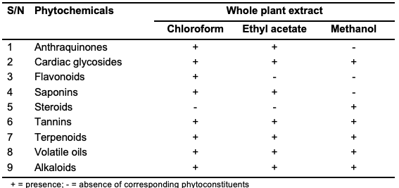
Results and Discussion Phytochemical Screening The preliminary phytochemical screening of Jaundea pinnata indicates the presence of cardiac glycosides, tannins, terpenoids, volatile oils and alkaloids in all the three ex-tracts. Anthraquinones and saponins were both found in the chloroform and ethyl acetate extracts but not in the methanol extract. Flavonoids were found in the chloroform extract only, whereas steroids were found to be present in the methanol extract only (Table 1). The presence of this important class of phytochemicals could be an indicator that Jaundea pinnata may have pharmacological importance. For instance, as natural polyphenols, flavonoids are well-recognized for their exceptional antioxidant properties [16,18]. Concurrently, tannins, such as condensed tannins, fall under the category of phenolic compounds, characterized by the presence of oligomers and polymers of flavan-3-ol monomeric units. Numerous studies have reported diverse biological activities associated with condensed tannins, including noteworthy antioxidant effects [16,19-21].
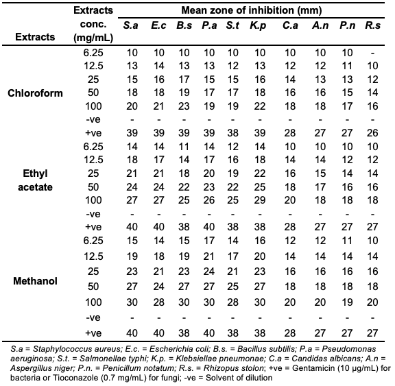
Antimicrobial Activity The antimicrobial assay is presented in Table 2. From the antibacterial study, the chloroform, ethyl acetate and methanol extracts inhibited the growth of all the test organisms at all concentrations, although the test organisms were just fairly inhibited at low concentrations (6.25–100 mg/mL). At moderate to high concentrations (50–100 mg/mL), all the three extracts showed significant inhibition of all the test organisms. The methanolic extract showed better inhibition than the ethyl acetate extract, whereas the chloroform extract displayed the least inhibition of the test organisms. However, the results of the antifungal assay indicated effective inhibition of all the test organisms by all the three extracts, at all concentrations, except for the chloroform extract which was resisted by Rhizopus stolonifer at a concentration of 6.25 mg/mL. Here, the inhibition also increases with increase in the concentration. The test organisms were only fairly inhibited at lower concentrations, between 6.25 and 25 mg/mL, but at medium to high (50–100 mg/mL), the extracts showed significant antifungal activity. In addition, the methanol extract displayed higher anti-fungal activity than the chloroform extract although with almost similar activity as the ethyl acetate extract.
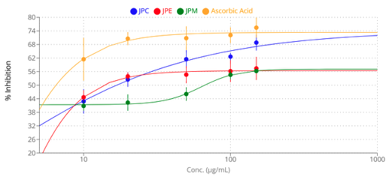
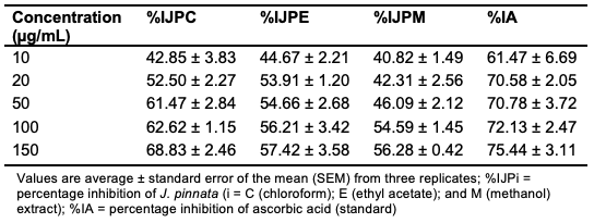
From the foregoing, it can be deduced that the three extracts exhibit antimicrobial activity and their effects on the microbes depend on the concentration of the extract – the higher the concentration, the higher the antimicrobial activity. The activity may be attributed to the presence of bioactive compounds, such as terpenoids, alkaloids and tannins. The better inhibition of methanol extract, compared to the chloroform and ethyl acetate extracts, may be due to the presence of steroids, as shown in Table 1. Antioxidant Activity The reduction ability of DPPH radical, indicative of antioxidant activity, was determined by measuring the decrease in absorbance induced by plant antioxidants– a process wherein the free radical accepts an electron or hydrogen radical, transforming into a stable diamagnetic molecule [16]. Hence, the scavenging potential of the plant extracts (chloroform, ethyl acetate, and methanol) against DPPH radicals were analyzed,and the results of the analysis are shown in Table 3 and Figure 1.
The results indicated that the extracts scavenged DPPH radicals in a dose-dependent manner with the chloroform extract displaying the highest percentage inhibition (68.83%) at 150 µg/mL.
Illustrated in Figure 1 are the scavenging effects of the three plant extracts and the standard on DPPH radicals, with their IC50 values determined using an IC50 calculator [22], based on a dose-dependent, non-linear regression algorithm. The IC50 values of chloroform, ethyl acetate, and methanol extracts are 2.11 µg/mL, 3.16 µg/mL, and 63.25 µg/mL, respectively, whereas that of ascorbic acid is 2.42 µg/mL. The observed IC50 values indicate pronounced antioxidant properties for both the chloroform and ethyl acetate extracts of the plant, comparable to those of the reference, ascorbic acid. Notably, considering the established inverse relationship between antioxidant activity and IC50 values [16], the chloroform extract of J. pinnata exhibited the highest antioxidant activity (greater than the reference) among the tested extracts while the methanol extract displayed the weakest antioxidant property. These results can be attributed to the presence of flavonoids in the chloroform extract, which were found lacking in the remaining extracts (Table 1). Despite the absence of flavonoids in both the ethyl acetate and methanol extracts, as revealed by the results from the phytochemical screening (Table 1), the IC50 value of the former was very much lower than that of the latter. Possible explanations for this phenomenon may be attributed to the presence of condensed tannins in the ethyl acetate extract, which could be lacking or present in a trace quantity in the methanol extract, or the presence of certain anthraquinones (such as emodin, aloe-emodin, chrysophanol, physcion, rhein, and hypericin) in the ethyl acetate extract, which are known for their reported antioxidant activity [23]. Nevertheless, a more detailed phytochemical profiling, including total phenolic content and anthraquinone profiling, is warranted to substantiate these hypotheses.
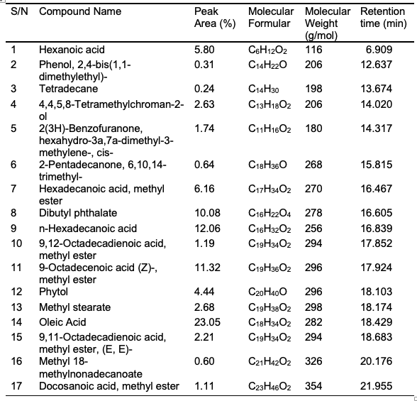
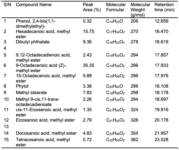
GC-MS Results Shown in Tables 4-6 are the GC-MS results from the chloroform, ethyl acetate, and methanol extracts of J. pinnata (aerial parts). In the chloroform extract, 10 compounds were identified, the principal one being phytol (5.60%). In contrast, the ethyl acetate extract revealed 17 compounds. Its primary constituents comprised oleic acid (23.05%), n-hexadecanoic acid (12.06%), methyl ester of 9-octadecenoic acid (Z) (11.32%), dibutyl phthalate (10.08%), hexadecanoic acid methyl ester (6.16%), and hexanoic acid (5.80%). Conversely, 15 compounds were identified in the methanol extract, the principal constituents being methyl ester of 9-octadecenoic acid (Z) (35.35%), hexadecanoic acid methyl ester (15.75%), dibutyl phthalate (9.36%), methyl stearate (7.83%), and 15-octadecenoic acid methyl ester (5.89%). These principal components, along with the trace compounds, are likely responsible for the observed free radical scavenging and antimicrobial activities of the plant extracts.
Phytol, present at 5.60% in the chloroform extract (Table 4) has been reported for its potent in vitro antioxidant activity, attributed to its potential to eliminate hydroxyl radicals and nitric oxide and prevent the formation of thiobarbituric acid reactive substances [24]. Its presence, together with the existence of flavonoids (Table 1), may elucidate the chloroform extract’s comparatively higher antioxidant property, despite its comparatively lower antibacterial and antifungal activities among the three extracts. Detailed compound information, including compound names, peak areas, molecular formulae, molecular weights and retention times, is provided in Tables 4, 5, and 6 for all the constituents present in each extract. The compounds reported in the table are the ones with highest matching (> 95%) of spectra and fragmentation patterns with reference to those from the NIST’s mass spectral library.
Conclusions and Recommendation The aerial parts of Jaundea pinnata have been investigated in this research and the preliminary phytochemical assay revealed the presence of cardiac glycosides, tannins, terpenoids, volatile oils and alkaloids in all the three extracts. Notably, the absence of flavonoids in the ethyl acetate and methanol extracts, steroids in the chloroform and ethyl acetate extracts, and anthraquinones solely in the methanol extract was also observed. Significant antimicrobial activity was noted across all the three extracts, demonstrating notable inhibition of all the test bacteria and fungi at moderate to high concentrations (50-100 mg/mL). Although relatively lower than those exhibited by the positive control (Table 2: Gentamicin for bacteria or Tioconazole for fungi), the dose-dependent nature of the extracts’ activity suggests the possibility to match the antimicrobial activity of the positive control at higher concentrations, say ≥ 200 mg/mL. This supports the ethnomedicinal use of the plant in traditional medicine for conditions associated with bacteria and fungi infections. Furthermore, the plant extracts exhibited substantial efficacy in scavenging DPPH radicals comparable to that of the reference, ascorbic acid, with the chloroform extract displaying slightly higher antioxidant property, thereby affirming their potential therapeutic utility in oxidative stress-related diseases. Gas Chromatography-Mass Spectrometry (GC-MS) analysis of the extracts identified several peaks corresponding to bioactive compounds, including phytol and oleic acid, which can act as free radical scavengers, helping to neutralize reactive oxygen species (ROS) and reduce oxidative stress in the body. This comprehensive research, encompassing phytochemical screening, radical scavenging activities, antimicrobial properties, and GC-MS analysis, collectively underscores the rationale for considering the utilization of this plant in alternative and traditional medicine. The study highlights its potential efficacy in treating various ailments, especially those associated with oxidative stress, injury, or microbial infections. To this end, further investigations are recommended to achieve a comprehensive isolation, identification, characterization, and elucidation of the structures of the bio-active compounds responsible for the observed pharmacological effects. The possibility of the extracts’ antimicrobial activities surpassing those of the positive controls at concentrations ≥ 150 mg/mL could also be verified as an extension of this study.
Funding The research received no external funding.
Conflict of Interest The authors declare no conflict of interest.
References
|
||||||||||||||||||||||||||||||||