The Chemist | Journal of the American Institute of Chemists
 
 
  TABLE OF CONTENTS
 
  EDITORIAL
 
  ARTICLE #1 -
  A COMPARISON OF
  TOTAL, FREE, & % FREE
  PROSTATE SPECIFIC
  ANTIGEN FOR THE
  SERODIAGNOSIS OF
  PROSTATE CANCER IN
  AFRICAN-AMERICAN &
  CAUCASIAN-AMERICAN
  MALES
 
  ARTICLE #2 -
  STUDIES ON
  CENOSPHERES AS
  FILLERS FOR PVC
  COMPOUNDS FOR
  APPLICATIONS IN
  ELECTRICAL CABLES
 
  ARTICLE #3 -
  COMPUTATIONAL
  STUDIES ON THE IR
  & NMR SPECTRA
  OF 2-AMINOPHENOL
 
  ARTICLE #4 -
  THE CHEMISTRY
  ASSOCIATED WITH AN
  EXPLODING LEAD-ACID
  BATTERY
 
  ARTICLE #5 -
  CHALLENGES FOR
  CHEMICAL EDUCATION:
  ENGAGING GREEN
  CHEMISTRY
  ENVIRONMENTAL
  SUSTAINABILITY
 
  ARTICLE #6 -
  CHEMISTRY
  EDUCATION:
  REFLECTION OF A
  MIDDLE SCHOOL
  TEACHER
 
  PUBLIC
  UNDERSTANDING
  OF CHEMISTRY -

  SCIENCE &
  TECHNOLOGY
  COMMUNICATION:
  NANOTECHNOLOGY - A
  POSITIVE ISSUE


The Chemist Volume 86 | Number 1 printDownload (pdf)
ISSN 1945-0702
 
Computational Studies on the IR and NMR Spectra of 2-Aminophenol
Abraham George1*, P, V, Thomas2, and David Devraj Kumar3
1. Department of Chemistry, Sun College of Engineering and Technology, Nagercoil, Erachakulam, Kanyakumari,
    TamilNadu-629 902, India
2. Thin Film Lab, Research Center, Post-Graduate Department of Physics, Mar Ivanios College,
    Thiruvananthapuram-695015, India
3. College of Education, Florida Atlantic University, 3200 College Avenue, Davie, FL 33314, USA (*mankavil.abraham@gmail.com)

 
Abstract: The geometry of 2-aminophenol has been optimized using HF and DFT methods using two basis sets 6-31 G(d) and cc-pVDZ using Gaussian 03 ™ software. The optimized geometry has been used for calculating the vibrational frequencies and that has been compared with the experimental values. The H1 and C13 NMR have been calculated by the Gauge Independent Atomic Orbitals (GIAO) method and have been found to be in good agreement with experimental values. The relative molecular orbital energies and shapes have been studied.

Key Words: Aminophenol, IR, NMR, GIAO, and Gaussian 03™.

Introduction

2-Aminophenol is an organic compound with molecular formula C6H4(OH)NH2. It is an amphoteric molecule and a reducing agent useful for the synthesis of dyes and heterocyclic compounds [1]. 2-Aminophenol can be prepared via the reduction of 2-nitrophenol, and it has a rather high melting point due to internal hydrogen bonding (m.p. 174 °C) [2].

A computational study on the enhanced stabilization of aminophenol derivatives by internal hydrogen bonding has been reported [3]. Jose et al. have conducted the density functional theory (DFT) study on the thermodynamic properties of aminophenols [4]. Experimental and theoretical investigation of first hyperpolarizability in aminophenols have been performed and reported by Franzen et al. [5].

In the present study, the vibrational and NMR spectra of the title compound have been calculated and compared with experimental results [10].

Computational Details

Calculations of the title compound were carried out with Gaussian 03™ software program [6]. The structure of 2-aminophenol has been optimized at two levels of theory using two basis sets. They are RHF/6-31 G (d), RHF/cc-pVDZ, B3LYP/6-31 G(d) and B3LYP/cc-pVDZ [7, 8]. Molecular geometries were fully optimized by Berny’s optimization algorithm using redundant internal coordinates. The structural parameters of 2-aminophenol determined by these calculations are listed in Tables 1 and 2 in accordance with the atom numbering scheme in Figure 1.

Tables 1 and 2 compare the calculated bond lengths and angles for 2-aminophenol with those of experimentally available data [10]. Using the optimized geometry, the vibrational frequencies were calculated using the same level of theory and the same basis sets. Harmonic vibrational wavenumbers were calculated using analytic second derivatives to confirm convergence to minima on the potential surface.

Table 1. Bond Lengths(Å) of 2-aminophenol
Atoms RHF/6-31G(d) RHF/cc-pVDZ B3LYP/6-31G(d) B3LYP/ cc-pVDZ

C1 - C2 1.39 1.39 1.41 1.41
C2 - C3 1.38 1.39 1.40 1.40
C3 - C4 1.38 1.39 1.40 1.40
C4 - C5 1.39 1.39 1.40 1.40
C5 - C6 1.38 1.38 1.40 1.40
C6 - C1 1.39 1.39 1.40 1.40
C6 - H15 1.07 1.08 1.09 1.09
C5 - H14 1.08 1.08 1.09 1.09
C4 - H13 1.07 1.08 1.09 1.09
C3 - H10 1.08 1.08 1.09 1.09
C2 - H11 1.43 1.43 1.45 1.45
N11 - H12 1.00 1.01 1.02 1.02
N11 - H9 1.00 1.01 1.02 1.02
C1 - O7 1.34 1.34 1.35 1.35
O7 - H8 0.95 0.95 0.98 0.98
 
Table 2. Bond Angles (o) of 2-aminophenol
Atoms RHF/6-31G(d) RHF/cc-pVDZ B3LYP/6-31G(d) B3LYP/ cc-pVDZ

C2 - C1 - C6 120.2 120.1 120.1 120.0
C2 - C1 - C7 120.5 120.5 119.4 119.2
C1 - C2 - C3 119.2 119.2 119.6 119.7
C1 - C2 - N11 116.5 116.5 114.9 114.8
C6 - C1 - O7 119.2 119.4 120.5 120.8
C1 - C6 - C5 119.7 119.8 119.4 119.5
C1 - C6 - H15 118.8 118.7 119.0 118.9
C1 - O7 - H8 107.3 106.9 103.9 103.4
C3 - C2 - N11 124.2 124.2 125.5 125.5
C2 - C3 - C4 120.9 121.0 120.4 120.4
C2 - C3 - H10 119.0 118.9 119.2 119.2
C2 - N11 - H9 119.9 111.3 111.4 110.9
C2 - N11 - H12 119.9 111.3 111.4 110.9
C4 - C3 - H10 120.1 120.1 120.4 120.4
C3 - C4 - C5 119.2 119.2 119.5 119.5
C3 - C4 - H13 120.3 120.3 120.1 120.2
C5 - C4 - H13 120.5 120.5 120.4 120.4
C4 - C5 - C6 120.7 120.7 120.9 120.9
C4 - C5 - H14 119.9 120.0 119.8 119.8
C6 - C5 - H14 119.4 119.4 119.3 119.3
C5 - C6 - H15 121.5 121.5 121.6 121.6
C2 - N11 - H12 107.8 107.0 107.0 106.0

At the optimized structure (Figure 1) of the examined species, no imaginary wave number modes were obtained, proving that a true minimum on the potential surface was found. The proton NMR and C13 NMR of the title compound have been calculated using the gauge independent atomic orbitals (GIAO) method.

Results & Discussion

Bond Length

All C-C bond lengths in 2-aminophenol have the same value in both HF and DFT methods. The C-H bond length exhibits slight variations when basis set changes from 6-31G (d) to cc-pVDZ in the Hartee-Fock (HF) method. The same bond length has unaltered values in the DFT method. N-H bond length has the same values in DFT method for both basis sets, whereas in HF method bond length increases by 0.01Å as the basis set changes from 6-31G(d) to cc-pVDZ. C=O bond length has same values in the HF and DFT methods. The C=O bond length increases by 0.01Å as the theory changes from RHF to DFT. O-H bond length has same values in HF and DFT method as basis set varies from 6-31G(d) to cc-pVDZ. O-H bond length increases by 0.03Å as theory changes from RHF to DFT. The bond lengths of 2-amino phenol are given in Table 1.

Bond Angles

In 2-aminophenol the bond angle values obtained by RHF and B3LYP calculations exhibit a slight difference in 6-31G(d) and cc-pVDZ basis sets. But in some cases, like C1-C2-N11, C1-O7-H8, C6-C1-O7, C4-C5-H14, there is much difference in bond angles of RHF and B3LYP calculations. The bond angles of 2-amino phenol are given in Table 2.

Vibrational Frequencies

The vibrational frequencies were calculated computationally using two basis sets 6-31G(d) and cc-pVDZ and two theories RHF and B3LYP. The DFT hybrid B3LYP tends to overestimate the fundamental modes, therefore scaling factors have to be used for obtaining a considerably better agreement with experimental data. Therefore, a scaling factor of 0.9613 and 0.8929 was uniformly applied to the wavenumbers calculated using DFT and HF, as suggested in Gaussian 03™ [9]. The vibrational frequency obtained from the calculation using RHF method was multiplied by a factor 0.8929 and by 0.9613 in DFT method for scaling. The frequencies are then being compared with the experimental spectrum obtained from the SDBS website [10]. The computational and experimental frequencies of 2-aminophenol with their assignments are given in Table 3 [11, 12].

Table 3. Infra Red Vibrational Frequencies(cm-1) and their assignments
Modes of Vibration RHF/6-31G(d) RHF/cc-pVDZ B3LYP/6-31G(d) B3LYP/ cc-pVDZ

  N-H assym.stretching 3412 3386 3404 3390
  O-H stretching 3378 3620 3584 3393
  C-H sym. stretching 3093 3013 3031 3080
  C-H assym. stretching 3082 3004 3021 3072
  C-H assym. stretching 3064 2985 3003 3054
  C-H assym. stretching 3051 2973 2990 3041
  N-H bending 1626 1608 1636 1642

H1 NMR

The proton NMR of the title compound has been calculated using the gauge independent atomic orbitals (GIAO) method [13, 14]. From the isotropic values of the molecule and that of TMS we obtain the δ values of the nmr spectrum. The isotropic value of the protons of TMS is 31.6198 in the RHF calculation and 31.7825 in B3LYP calculation. The proton-nmr spectrum of 2-aminophenol is taken from the website SDBS [10]. The δ values of 2-aminophenol are given in Table 4.

Table 4. δ values of H1 NMR of 2-aminophenol
Proton RHF/6-31G(d) RHF/cc-pVDZ B3LYP/6-31G(d) B3LYP/cc-pVDZ Exeperimental Value [10]

  8 4.5343 5.4842 6.9493 7.4618 8.9
  9 & 12 0.0959 1.2265 1.2968 1.9253 4.4
0.0952 1.2272 1.2977 1.9262 4.4
  15 5.9322 7.0589 6.6391 7.0936 6.587
  10 5.4016 6.5323 6.2952 6.7283 6.543
  13 6.0264 7.1094 6.7479 7.1718 6.649
  14 5.670 6.7859 6.4479 6.8969 6.400

The proton-nmr spectrum of 2-aminophenol exhibits six peaks. This indicates six types of protons. The computationally calculated values of protons 8, 9, 12, 10, 15, 13 and 14 agree with the experimental values, but the computationally calculated values of protons 9 and 12 are less than the experimental values in both HF and DFT methods. The chemical shift values calculated by GIAO method show slight difference from the experimental values, but maintain a general trend of chemical shift.

C13 NMR

The isotropic value of the C13 of TMS is 195.1196 in the RHF theory and 188.7879 in the theory B3LYP. The C13 nmr spectrum of the molecule is taken from the website SDBS [10]. From the isotropic values of the carbon of 2-aminophenol and the TMS we obtain the δ values of their C13 nmr spectrum. The δ values of 2-aminophenol are given in Table 5. The C13 spectrum of 2-aminophenol reveals 6 peaks, which indicates 6 different types of carbon atoms. From the computational calculations also we have 6 values which agree with the experimental values.

Table 5. δ values of C13NMR of 2-aminophenol
C13 type RHF/6-31G(d) RHF/cc-pVDZ B3LYP/6-31G(d) B3LYP/cc-pVDZ Exeperimental Value [10]

1 146.123 149.46 151.84 143.13 143.95
2 114.21 117.84 122.64 111.51 116.48
3 123.22 125.91 123.52 119.58 136.40
4 107.86 110.04 112.48 103.71 114.47
5 122.64 125.12 123.65 118.79 119.47
6 106.04 107.86 109.13 101.53 114.44

HOMO-LUMO energy gaps

The relative energy of the molecular orbitals have been calculated and a graphical representation of the highest occupied molecular orbital (HOMO) and lowest unoccupied molecular orbital (LUMO) of 2-aminophenol are given in Figures 2(a) and (b).

Figure 2(a). HOMO of 2-aminophenol | Figure 2(b). LUMO of 2-aminophenol

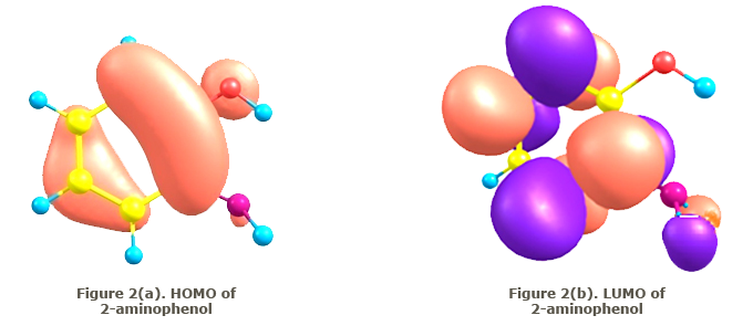
Conclusion

The structure of 2-aminophenol was optimized by the RHF and DFT methods using the basis sets 6-31 G (d) and cc-pVDZ. Using the optimized geometry, the vibrational frequencies, proton NMR and C13 NMR of the title compound have been calculated and have been found to agree well with experimentally reported values. The small differences are due to the fact that experimental values are recorded in the solid state and theoretical calculations belong to the gaseous phase. A plot of the highest occupied molecular orbital (HOMO) and that of the lowest unoccupied molecular orbital (LUMO) is also made.

References

  1. Mitchell SC ,Waring RH in “Aminophenols:” Ullmann’s Encyclopedia of Industrial Chemistry, Wiley-VCH, 2002.
  2. Reference Handbook of Fine Chemicals, Acros Organics Publishers, Fisher Scientific UK, 2007.
  3. Gomes JRB, Manuel AV, da Silva R. Chemical Physics, 2006, 324, 600.
  4. Gomes JRB, Manuel AV, Da Silva R. International Journal of Quantum Chemistry, 2005, 101, 860.
  5. Franzen PL, Zilio SC, Machado AEH, Madurro JM, Brito-Madurro AG, Ueno LT, Sampaioand RN, Barbosa Neto NM. J. Mol. Structure, 2008, 892, 254.
  6. Gaussian 03, Frisch MJ, Trucks GW, Schlegel HB, Scuseria GE, Robb MA, Cheeseman JR, Montgomery, JA, Jr., Vreven T, Kudin KN, Burant JC, Millam JM, Iyengar SS, Tomasi J, Barone V, Mennucci B, Cossi M, Scalmani G, Rega N, Petersson GA, Nakatsuji H, Hada M, Ehara M, Toyota K, Fukuda R, Hasegawa J, Ishida M, Nakajima T, Honda Y, Kitao O, Nakai H, Klene M, Li X, Knox JE, Hratchian HP, Cross JB, Bakken V, Adamo C, Jaramillo J, Gomperts R, Stratmann RE, Yazyev O, Austin AJ, Cammi R, Pomelli C, Ochterski JW, Ayala PY, Morokuma K, Voth GA, Salvador P, Dannenberg JJ, Zakrzewski VG, Dapprich S, Daniels AD, Strain MC, Farkas O, Malick DK, Rabuck AD, Raghavachari K, Foresman JB, Ortiz JV, Cui Q, Baboul AG, Clifford S, Cioslowski J, Stefanov BB, Liu G, Liashenko A, Piskorz P, Komaromi I, Martin RL, Fox DJ, Keith T, Al-Laham MA, Peng CY, Nanayakkara A, Challacombe M, Gill PMW, Johnson B, Chen W, Wong M.W, Gonzalez C, Pople JA, Gaussian, Inc., Wallingford, CT.
  7. Becke AD. J. Chem. Phys, 1993, 98, 5648.
  8. Lee C, Yang W, Parr RG. Phy. Rev., 1998, B37,785.
  9. Foresman JB in Exploring Chemistry with Electronic Structure Methods: A Guide to using Gaussian, ed. E. Frisch, Pittsburg, PA, 1996.
  10. SDBSWeb : http://riodb01.ibase.aist.go.jp/sdbs/ (National Institute of Advanced Industrial Science and Technology, 06, August, 2012)
  11. Socrates G in Infrared Characteristic Group Frequencies, John Wiley and Sons, New York, 1980.
  12. Coates J, Meyers RA in Interpretation of Infrared Spectra, A Practical Approach, Chichester, John Wiley and Sons Ltd., 2000.
  13. Szelejewska-Woźniakowska A, Chilmonczyk Z, Leś A, Cybulski J, Wawer, I. Solid State Nuclear Magnetic Resonance, 1999, 14(1), 59
  14. Wolinsky K, Hilton JF, Pulay P. J. Am. Chem. Soc., 1990, 112.


All rights are reserved. © The AIC 2013.
The American Institute of Chemists, Inc. does not necessarily endorse any of the facts or opinions expressed in the articles, book reviews, or advertisements appearing in The Chemist.

Web site designed and developed by Maxide Roson, LLC

The American Institute of Chemists, Inc.
315 Chestnut Street, Philadelphia, PA 19106-2702.
Phone (215) 873-8224 | Fax: (215) 629-5224
E-mail: aicoffice@TheAIC.org